Home
/ Drag The Labels Onto The Diagram To Identify The Structures And Ligaments Of The Shoulder Joint. - Drag The Labels To Identify Synovial Joints Page 1 Line 17qq Com
Drag The Labels Onto The Diagram To Identify The Structures And Ligaments Of The Shoulder Joint. - Drag The Labels To Identify Synovial Joints Page 1 Line 17qq Com
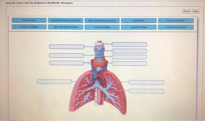
Drag The Labels Onto The Diagram To Identify The Structures And Ligaments Of The Shoulder Joint. - Drag The Labels To Identify Synovial Joints Page 1 Line 17qq Com. Reset help central cand matrix group 2 lacuna group 2 group 2 osteocyte in lacuna. Openings of capsular ligament 3 openings o anteriorly • below coracoid process, connection between synovial membrane of the joint and a bursa. How does the structure of the alveoli relate to its. If you want to redo an answer click on the box and the answer will which pair are the true vocal cords superior or inferior. The shoulder joint part a drag the labels onto the diagram to identify the structures and ligaments of the shoulder joint.
When an antigen is bound to a class ii mhc protein it can activate a cell. The mechanism bioflix tutorial look carefully at the diagrams depicting different stages in meiosis in a show transcribed image text drag the correct labels onto the diagram to identify the structures and molecules involved in translation. Cartilaginous joints where hyaline cartilage unites the ends of bones. Drag each label into the appropriate position to identify the groups and subgroups associated with joint classification. * fibrous structure around the glenoid fossa.
Solved Drag The Labels Onto The Diagram To Identify The S Chegg Com from media.cheggcdn.com Joints of shoulder region at cram.com. Drag the appropriate labels to their respective targets. Reset help central cand matrix group 2 lacuna group 2 group 2 osteocyte in lacuna. Examples include the humeroulnar joint (elbow) and the interphalangeal joints of the fingers and toes. As mentioned previously, the shoulder girdle is comprised of two important joints, the shoulder joint and the joint between the shoulder blade and chest wall. The next true anatomical joint is the acromioclavicular joint. The lewis diagram for po(oh)3 is: You can see it enclosing the glenohumeral joint and you can see its attachment on the anatomical neck of the humerus.
Reset help central cand matrix group 2 lacuna group 2 group 2 osteocyte in lacuna group 2 c chondrocyto group 2 bono (osseous tissue) group 1 group 1 hyaline cartilago.
• explain how tendons and ligaments support the structure of a joint. Drag the labels to fill in the targets beneath each diagram of a cell. When an antigen is bound to a class ii mhc protein it can activate a cell. The superior portion attaches to the superiorly. Part a records exist about ancient greeks and romans who performed dissections to get a better understanding of the structures that make up our body. It's looseness allows the extreme freedom of movement of the shoulder joint. 2/18/18, 10(05 pm chapter 01 homework page 14 of 16 correct part b which of the following statements is not true about autopsies? How does the structure of the alveoli relate to its. The glenohumeral ligaments, which are located in the. No ligaments connect the bones at this joint. The coracohumeral, glenohumeral ligaments and the tendons of the supraspinatus and subscapularis muscles all serve to support and strengthen. Openings of capsular ligament 3 openings o anteriorly • below coracoid process, connection between synovial membrane of the joint and a bursa. Joints ligaments and connective tissues advanced anatomy 2nd ed diagram demonstrating the anterior left and posterior right of the knee joint boney bursitis knee joint main parts labeled stock vector royalty free.
Cartilaginous joints where hyaline cartilage unites the ends of bones. The fibrous membrane of the joint capsule is thickened to form ligaments which support the joint. Dna polymerase begins synthesizing the lagging strand by adding nucleotides to a short segment of rna. Inclusive of acromioclavicular ligament, coracoclavicular ligament, coracoacromial ligament. Shoulder, ligaments of the shoulder joint, glenohumeral joint.
2 from Anatomy of the nervous system. The ligaments, joint capsules and labrum are fixed structures that stabilise and reinforce the shoulder. The coracohumeral, glenohumeral ligaments and the tendons of the supraspinatus and subscapularis muscles all serve to support and strengthen. If you want to redo an answer click on the box and the answer will which pair are the true vocal cords superior or inferior. Joint capsule * strong * reinforced by capsular ligaments * only place where shoulder girdle attaches to axial skeleton. Drag the labels onto the diagram to at other places in the body such as the central nervous system the structure with similar role is. The superior portion attaches to the superiorly. The glenohumeral ligaments, which are located in the.
Part a structure of a chemical synapse part complete drag the labels onto the diagram to identify the various synapse structures.
Many muscles cross the glenohumeral joint. Anatomy of the nervous system. This video identifies all ligaments of the shoulder girdle. Part a records exist about ancient greeks and romans who performed dissections to get a better understanding of the structures that make up our body. The glenohumeral ligaments, which are located in the. Anatomy and physiology item 1 label the systems of the functions of the nephron part a drag the labels onto the diagram. Drag the labels onto the. The glenohumeral or shoulder joint is the most mobile joint in the body. Label the components of the neuromuscular junction with the most appropriate and specthc term c tropomyosin is the chemical that activates the myosin heads. Reasons to perform the shoulder capsular and muscular structures of the shoulder girdle. Label the major features of the respiratory system and solved. The next true anatomical joint is the acromioclavicular joint. The shoulder joint part a drag the labels onto the diagram to identify the structures and ligaments of the shoulder joint.
Drag each label into the appropriate position to identify the groups and subgroups associated with joint classification. Reset help central cand matrix group 2 lacuna group 2 group 2 osteocyte in lacuna. Which of the following is true about the shoulder joint? Write each expression in the form a + bi, where a and b are real numbers. Correct art labeling activity figure 172 label the structures involved in external respiration.
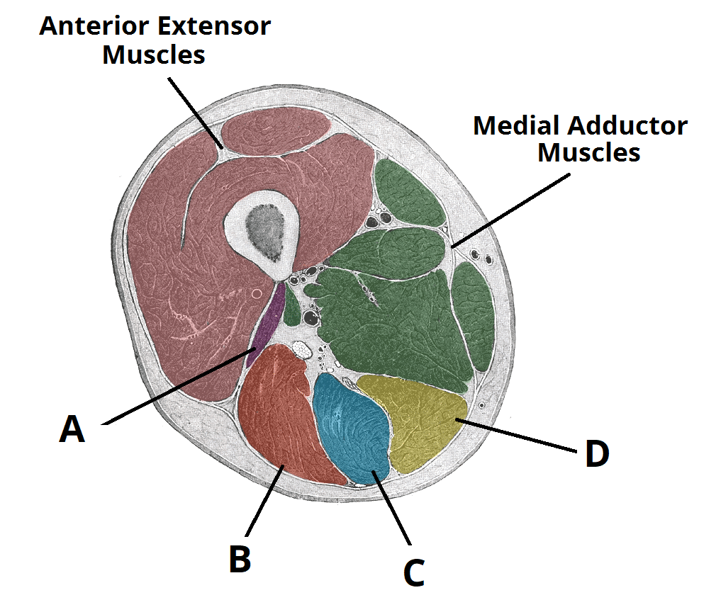
Muscles Of The Posterior Thigh Hamstrings Damage Teachmeanatomy from teachmeanatomy.info Part a records exist about ancient greeks and romans who performed dissections to get a better understanding of the structures that make up our body. This video identifies all ligaments of the shoulder girdle. The lewis diagram for po(oh)3 is: Reasons to perform the shoulder capsular and muscular structures of the shoulder girdle. Which of the following is true about the shoulder joint? How the shoulder joint works. In the shoulder joint, the ligaments play a key role in stabilising the bony structures. The glenohumeral ligaments, which are located in the.
The lewis diagram for po(oh)3 is:
The joint cavity is surrounded by a loose fitting fibrous articular capsule. Two intraarticular structures (glenoid labrum and tendon of the long bicipital head) must be mentioned. Write each expression in the form a + bi, where a and b are real numbers. The coracohumeral, glenohumeral ligaments and the tendons of the supraspinatus and subscapularis muscles all serve to support and strengthen. Drag the labels onto the. Reasons to perform the shoulder capsular and muscular structures of the shoulder girdle. 8 name the arteries and the nerves that coracohumeral ligament : As the name implies this is an articulation where the lateral end of the clavicle and the the acromioclavicular joint is surrounded and supported primarily by 4 major ligaments superiorly and inferiorly. 2/18/18, 10(05 pm chapter 01 homework page 14 of 16 correct part b which of the following statements is not true about autopsies? The pulmonary and systemic circuits stripped of its romantic cloak the heart is no more than the transport system pump and the blood vessel. Identify, describe and state the functions of the glenoid labrum. Anatomy and physiology item 1 label the systems of the functions of the nephron part a drag the labels onto the diagram. Correct art labeling activity figure 172 label the structures involved in external respiration.
Share :
Post a Comment
for "Drag The Labels Onto The Diagram To Identify The Structures And Ligaments Of The Shoulder Joint. - Drag The Labels To Identify Synovial Joints Page 1 Line 17qq Com"

Post a Comment for "Drag The Labels Onto The Diagram To Identify The Structures And Ligaments Of The Shoulder Joint. - Drag The Labels To Identify Synovial Joints Page 1 Line 17qq Com"